|

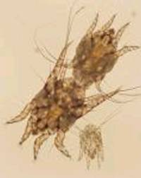
Ear mites are an external parasite that can infect dogs, cats and more rarely, humans. The mite's name
is Otodectes cynotis. Otodectes mites are large enough that they can sometimes be seen with the naked eye and are easy to
see with magnification.
Ear mites live in the ears and on the skin of pets infected with them. The mite lives its entire life on the pet and it
takes about 3 weeks for a mite egg to develop into an adult mite. The adult mites are very mobile and can live for some time
off of a dog or cat, which enables it to be fairly contagious.
The most common sign of ear mite infection is shaking of the head and ears. Dogs may also scratch at their ears, rub their
face and in severe cases may even cause bleeding sores behind their ears in their effort to relieve the discomfort from the
mites. The intense itching associated with these mites is thought to be due to a hypersensitivity reaction, which is similar
to an allergy. Some pets can be infected without showing signs of itching or head shaking, apparently because they don't develop
the hypersensivity reaction.
Ear mites are more commonly diagnosed in cats than they are in dogs but they
are a significant cause of ear infections
in dogs, too. Dark brown to black debris accumulates in the ears of infected pets and the mites may be visible as small moving
white specks on the debris. Secondary infection with bacteria or yeast is common in ear mite infections and may complicate
the diagnosis. The mites can live on the skin and some dogs and cats appear to have infections that affect only the skin,
causing small sores to develop in affected areas. It is important to treat the ears for mites and the whole pet with a product
that is capable of killing the mites. Most flea and tick products will kill ear mites on the skin.
In multiple pet households it is important to treat all the pets and to clean the environment, considering the use of premise
control insecticides in persistent cases. Ear mites are susceptible to many medications, including pyrethrins, rotenone, fibronil,
thiabendazole and ivermectins. It is necessary to treat for at least three to four weeks in most instances to be sure to kill
the adult mites and any eggs that may hatch later.
Many veterinary clients treat their dog's ears with over the counter products for ear mites based on the presence of ear
inflammation or exudate in the ears, doing this for weeks or months prior to giving up and having their dog's ears examined.
There are a number of causes of ear infection in dogs and it is best to have your vet examine your dog's ears to determine
if the cause of ear irritation is ear mites or another infection. Doing this can save your dog from weeks of pain or discomfort.
Mike Richards, DVM
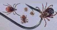
Dogs get ehrlichiosis from the brown dog tick, which passes an Ehrlichia organism into the bloodstream
when it bites. There are three stages of ehrlichiosis, each varying in severity. The acute stage, occurring several weeks
after infection and lasting for up to a month, can lead to fever and disorders of the blood. The second stage, called the
subclinical phase, has no outward signs and can last for up to five years. If the infected dog’s immune system is unable
to eliminate the Ehrlichia organism, the third and most serious stage of infection, the chronic phase, will commence.
Lameness, neurological and ophthalmic disorders, kidney disease, and anemia and other blood disorders can result. Chronic
ehrlichiosis can be fatal.
Antibiotics, administered for an extended period of time, are effective at eliminating the infection. Dogs
with severe cases of chronic ehrlichiosis cannot be cured, but supportive care and treatment of diseases secondary to the
infection, such as anemia, can help stabilize the dog.
| Canine Hip Dysplasia |
| |
|
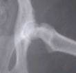
Hip dysplasia is a genetic disorder in which dogs have a poor fitting hip joint. This ball and socket joint
should fit together neatly, allowing dogs to move the legs freely and without pain. Because their bones do not fit properly,
dogs with hip dysplasia are prone to develop arthritis and related joint pain as they age. Motion of the hip joints slowly
causes erosion of soft cartilage in these joints. Hip dysplasia can affect either or both of the rear leg joints.
Hip dysplasia can occur in most breeds, but it is predominant in larger dogs, particularly the German Shepherd,
St. Bernard, Labrador Retriever, Pointers, and Setters. Although hip dysplasia is a genetic condition, research shows that
environmental factors can also put a dog at risk. Overfeeding (especially of puppies) can predispose a dog to hip dysplasia.
Excessive exercise may predispose dogs as well.
Diagnosis
Signs of severe hip dysplasia usually appear
before the dog reaches one year of age. Signs include rear leg pain, incoordination, and a limp or wavering gait. A common
sign is the dog that has trouble rising. Dogs with severe hip dysplasia typically develop lameness by two years of age. Dogs
with less severe cases may not experience arthritis and the related pain or lameness until six to ten years of age.
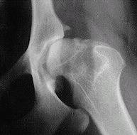
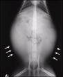
Since the clinical signs of hip dysplasia are similar to those of other diseases, veterinarians rely on X-rays
to make a final diagnosis. This requires a mild anesthetic in order to carefully position the dog on the radiographic table.
Veterinarians look for degenerative changes and abnormal shapes of the hip joint.
Treatment
Depending on the severity of the dog's condition,
veterinarians treat hip dysplasia with either drugs or surgery. Drug therapy doesn't reverse or cure the progression of hip
dysplasia, but it does offer relief from the associated pain. There are several steroidal and non-steroidal, anti-inflammatory
drugs available through veterinarians. Most require daily administration. For many dogs, these prescriptions can offer a tremendous
relief--they return to a more active lifestyle that is free of joint pain. | |
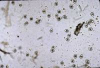
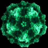
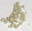
What are Coccidia?
Coccidia are single celled organisms that infect the intestine. They are microscopic parasites detectable on
routine fecal tests in the same way that worms are but coccidia are not worms and they are not visible to the naked eye. Coccidia
infection causes a watery diarrhea which is sometimes bloody and can even be a life-threatening problem to an especially young
or small pet.
Where do Coccidia Come From?
Oocysts (pronounced o'o-sists), like those shown above, are passed in stool. In the outside world, the oocysts
begin to mature or “sporulate.” After they have adequately matured, they become infective to any host (dog or
cat) that accidentally swallows them.
To be more precise, coccidia come from fecal-contaminated ground. They are swallowed when a pet grooms/licks the dirt off
itself. In some cases, sporulated oocysts are swallowed by mice and the host is infected when it eats the mouse. Coccidia
infection is especially common in young animals housed in groups (in shelters, rescue areas, kennels, etc.) This is a common
parasite and is not necessarily a sign of poor husbandry.
What Happens Inside the Host?
The sporulated oocyst breaks open and releases eight sporozoites. These sporozoites each finds an intestinal
cell and begins to reproduce inside it. Ultimately, the cell is so full of what are called “merozoites” that it
bursts releasing the merozoites which seek out their own intestinal cells and the process begins again. It is important to
note how thousands of intestinal cells can become infected and destroyed as a result of accidentally swallowing a single oocyst.
As the intestinal cells are destroyed in larger and larger numbers, intestinal function is disrupted and a bloody, watery
diarrhea results. The fluid loss can be dangerously dehydrating to a very young or small pet.
How Are Coccidia Detected?
A routine fecal test is a good idea for any new puppy or kitten whether there are signs of diarrhea or not as
youngsters are commonly parasitized. This sort of test is also a good idea for any patient with diarrhea. The above illustration
demonstrates coccidia oocysts seen under the microscope in a fecal sample. Coccidia are microscopic and a test such as this
is necessary to rule them in. It should be noted that small numbers of coccidia can be hard to detect so just because
a fecal sample tests negative, this does not mean that the pet is not infected. Sometimes several fecal tests are performed,
especially in a young pet with a refractory diarrhea; parasites may not be evident until later in the course of the condition.
How is Coccidiosis Treated?
We do not have any medicine that will kill coccidia; only the patient’s immune system can do that. But
we can give medicines called “coccidiostats” which can inhibit coccidial reproduction. Once the numbers stop expanding,
it is easier for the patient’s immune system to “catch up” and wipe the infection out. This also means,
though, that the time it takes to clear the infection depends on how many coccidia organisms there are to start with and how
strong the patient’s immune system is. A typical treatment course lasts about a week or two but it is important to realize
that the medication should be given until the diarrhea resolves plus an extra couple of days. Medication should be given for
at least five days total. Sometimes courses as long as a month are needed.
The use of sulfa drugs in pregnancy can cause birth defects. Sulfa drug use can also lead to false positive test results
for urine glucose.
Can People or Other Pets Become Infected?
While there are species of coccidia that can infect people (Toxoplasma and Cryptosporidium, for
example), the Isospora species of dogs and cats are not infective to people. Other pets may become infected from exposure
to infected fecal matter but it is important to note that this is usually an infection of the young (i.e. the immature immune
system tends to let the coccidia infection reach large numbers where the mature immune system probably will not.) In most
cases, the infected new puppy or kitten does not infect the resident adult animal.

|
| Philodendron |
Poisonous Plants
Protect your furbaby - Use these links for names and photos of poisonous plants:
Pyometra, a serious infection of the uterus, is a well-recognized disease of female dogs. Pyometra often
results from the animal’s own bacteria within the genital tract. Escherichia coli is the most common bacteria identified
in pyometra. Whenever levels of the reproductive hormone progesterone rise, the uterine lining becomes susceptible to bacterial
infection.
Dogs with pyometra commonly have a vaginal discharge, fever, lethargy, and a loss of appetite. Affected
dogs are often dehydrated; some may drink and urinate excessively. Some dogs will appear asymptomatic until after vaginal
discharge begins. Others will go into shock. Laboratory tests often show dehydration-related abnormalities of electrolyte
balance and kidney function. Changes in the white blood cell count are common. Most patients are diagnosed using history,
clinical signs, physical examination, and abdominal x-rays.
Pyometra requires prompt treatment. Antibiotics to fight
the infection, and intravenous fluids to correct dehydration-related abnormalities, are routinely administered. Supportive
therapy is given to correct other organ system dysfunction and to stabilize the patient. Generally, surgical removal of the
uterus and ovaries is the preferred treatment.
Under construction...
|
 |
|
|
|
|
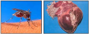
| One misquito can cause heartworms! |
Canine Heartworm Disease
Dogs are considered the definitive host for heartworms (Dirofilaria immitis). However, heartworms may infect more than 30 species of animals (e.g., coyotes, foxes, wolves and other wild canids, domestic
cats and wild felids, ferrets, sea lions, etc.) and humans as well. When a mosquito carrying infective heartworm larvae bites a dog and transmits the infection, the larvae grow, develop, and migrate in the body over a period of several months to become sexually mature male and female worms. These
reside in the heart, lungs, and associated blood vessels. Even as immature adults, the worms mate and the females release
their offspring (microfilariae), pronounced: (micro fil ar ee), into the blood stream. The time elapsed from when the larvae enter the dog until the minute offspring can be detected in the blood (pre-patent period) is about six to seven months. The
male heartworms (four to six inches in length) and the females (10-12 inches) become fully grown about one year after infection,
and their life span in dogs appears to be at least five to seven years.
In experimentally induced infections of heartworms in dogs, the percentage of infective larvae developing to adults is high (40% to 90%). However, the percentage of experimentally infected dogs from which adult worms
are recovered is virtually 100%. The number of worms infecting a dog is usually high, as the number of worms in dogs can range
from one to approximately 250.
Microfilaremia, the presence of heartworm offspring in the blood of the host, is relatively common in dogs. However, not all heartworm infections
result in such offspring circulating in the blood. These are known as occult heartworm infections and may be the result of a number of factors such as single sex heartworm infections, host immune responses
affecting the presence of circulating offspring (microfilariae) and most significantly, the administration of heartworm preventives.
The onset and severity of disease in the dog
is mainly a reflection of the number of adult heartworms present, the age of the infection, and the level of activity of the
dog. Dogs with higher numbers of worms are generally found to have more severe heart and lung disease changes. Until the number
of mature heartworms exceeds 50 in a 25-kg dog (approximately 55 pounds), nearly all of the heartworms reside in the lower
caudal pulmonary arteries (the arteries of the lower lung lobes). Higher numbers of heartworms will result in their presence in
the right chambers of the heart. In such infections, the most common early pathological changes caused by heartworms are due
to inflammatory processes that occur in and around the arteries of the lower portion of the lungs in response to the presence
of heartworms. Later, the heart may enlarge and become weakened due to an increased workload and congestive heart failure
may occur. A very active dog (e.g., working dog) is more likely to develop severe disease with a relatively small number of
heartworms than an inactive one (e.g., a lap dog or couch potato). In an occasional dog with a large number of heartworms,
the worms may not only be in the heart but also the caudal vena cava (large primary vein of the lower body) between the liver and the heart. This syndrome (Vena Cava or Liver Failure
Syndrome) is characterized by sudden collapse and even death within two to three days if they are not removed surgically.
Canine heartworm infection is widely distributed throughout the United States. Heartworm infection has been found in dogs
native to all 50 states. All dogs regardless of their age, sex, or habitat are susceptible to heartworm infection. The highest
infection rates (up to 45%) in dogs (not maintained on heartworm preventive) are observed within 150 miles of the Atlantic
and Gulf coasts from the Gulf of Mexico to New Jersey and along the Mississippi River and its major tributaries. Other areas
of the United States may have lower incidence rates (5% or less) of canine heartworm disease, while some regions have environmental,
mosquito, and dog population factors that allow a higher incidence of heartworm infection. Regions where heartworm disease
is common have infections diagnosed in dogs as young as one year of age, with most areas diagnosing infections primarily between
the ages of three and eight years. Although there are differences in frequency of infection for various groups of dogs, all
dogs in such regions should be considered at risk, placed on prevention programs and frequently examined by a veterinarian.
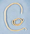
Roundworms
Roundworms are active in the intestines of puppies, often causing a pot-bellied appearance and poor growth. The worms may
be seen in vomit or stool; a severe infestation can cause death by intestinal blockage.
This worm can grow to seven inches in length. Females can produce 200 thousand eggs in a day, eggs that are protected by
a hard shell and can exist in the soil for years. Dogs become infected by ingesting worm eggs from contaminated soil. The
eggs hatch in the intestine and the resulting larva are carried to the lungs by the bloodstream.
The larva then crawls up the windpipe and gets swallowed, often causing the pup to cough or gag. Once the larvae return
to the intestine, they grow into adults.
Roundworms do not typically infest adults. However, as mentioned above, the larvae can encyst in body tissue of adult bitches
and activate during the last stages of pregnancy to infest puppies. Worming the bitch has no effect on the encysted larvae
and cannot prevent the worms from infecting the puppies.
Although roundworms can be treated with an over-the-counter wormer found in pet stores, a veterinarian is the best source
of information and medication to deal with intestinal parasites. Dewormers are poisonous to the worms and can make the dog
sick, especially if not used in proper dosage.
Kennel Cough-Tracheobronchitis
Canine infectious tracheobronchitis (kennel cough) is one of the most prevalent infectious diseases in dogs. Fortunately,
the majority of cases are not serious resolving on their own in 1 to 2 weeks . But because some dogs develop life- threatening
complications, you should take precautions to prevent your pooch from becoming infected with this highly contagious disease.
Kennel cough can be caused by a number of different airborne bacteria (such as Bordetella bronchiseptica) and viruses (such
as canine parainfluenza) or a mycoplasma (an organism somewhere between a virus and a bacteria). Typically, more than one
of these pathogens (disease-causing agents) must bombard the dog at once to trigger illness. Such a multifaceted attack is
most likely to occur when a dog spends time in close quarters with many other dogs. Dogs that attend dog shows, travel frequently,
or stay at kennels have a higher risk of developing kennel cough than do dogs that stay at home most of the time.
The primary sign of kennel cough is a dry- sounding, spasmodic cough caused by pathogens that
induce inflammation of the trachea (windpipe) and bronchi (air passages into the lungs). At the end of a coughing spell, a
dog will often retch and cough up a white foamy discharge. Some dogs also develop conjunctivitis (inflammation of the membrane
lining the eyelids), rhinitis (inflammation of the nasal mucous membrane), and a nasal discharge. Affected dogs usually remain
active and alert and continue to eat well. But if you suspect your dog has kennel cough, isolate it from other dogs and call
your veterinarian.
Parvovirus (Parvo)
Parvovirus is a viral disease of dogs. It affects puppies much more frequently than it affects adult dogs. The virus likes
to grow in rapidly dividing cells. The intestinal lining has the biggest concentration of rapidly dividing cells in a puppy's
body. The virus attacks and kills these cells, causing diarrhea (often bloody), depression and suppression of white blood
cells -- which come from another group of rapidly dividing cells. In very young puppies it can infect the heart muscle and
lead to "sudden" death.
Parvovirus Vaccination
Parvovirus is probably the most common viral illness of dogs at the present time. It is much more common in puppies than
it is in adult dogs. It can be very hard to successfully vaccinate a puppy for this disease because the antibody protection
the puppy acquires from its mother can interfere with vaccination. Many vets recommend vaccinating puppies every three to
four weeks for this virus starting at 6 weeks of age and continuing until they are at least 16 weeks of age and preferably
20 weeks of age. It is possible that this vaccine confers lifelong immunity once it does work but most veterinarians continue
to recommend yearly vaccination for it. It seems prudent to at least get the vaccination at one year of age. Since it is combined
with the other vaccines it is often easier just to give it yearly with them.
What are the symptoms of Parvo?
Parvo" is a virus that attacks the lining of the digestive system. It causes dogs and puppies to not be able to absorb
nutrients or liquids. Puppies are especially prone to it because they have an immature immune system. When dogs and puppies
contract parvo, they often have diarrhea, vomiting and lethargy. Usually they stop eating and develop a bloody, foul-smelling,
liquid stool.
Symptoms usually begin with a high fever, lethargy, depression, and loss of appetite. Secondary symptoms appear as severe
gastrointestinal distress, such as vomiting and bloody diarrhea. In many cases, dehydration, shock, and death follow.
Parvovirus is characterized by severe, bloody diarrhea and vomiting, high fever and lethargy. The diarrhea is particularly
foul smelling and is sometimes yellow in color. Parvo can also attack a dog's heart causing congestive heart failure. This
complication can occur months or years after an apparent recovery from the intestinal form of the disease. Puppies who survive
parvo infection usually remain somewhat un-healthy and weak for life.
How is Parvo transmitted?
Canine parvovirus is carried by dogs. Adult dogs may be infected carriers without showing any clinical signs. Dogs with
the typical diarrhea that parvovirus causes shed the virus as well. It can last a long time in the environment, perhaps as
long as 9 months or longer.
Generally, it takes 7-10 days from the time of exposure for dogs and puppies to start showing symptoms and to test positive
for parvo.
Parvo is highly contagious to unprotected dogs, and the virus can remain infectious in ground contaminated with fecal material
for five months or more if conditions are favorable. Extremely hardy, most disinfectants cannot kill the virus, however chlorine
bleach is the most effective and inexpensive agent that works, and is commonly used by veterinarians.
The ease with which infection with Parvo can occur in any unvaccinated dog must be stressed. The virus is extremely hardy
in the environment. Withstanding wide temperature fluctuations and most cleaning agents. Parvo can be brought home to your
dog on shoes, hands and even car tires. It can live for many months outside the animal. Any areas that are thought to be contaminated
with parvo should be thoroughly washed with chlorine bleach diluted 1 ounce per quart of water.
Dogs and puppies can contract parvo even if they never leave their yards. Parvo virus, despite what you might hear, is
NOT an airborne virus. It is excreted in the feces of infected dogs, and if someone -- human, dog, bird, etc. -- steps in
(or otherwise comes in contact with) the excrement, the possibility for contamination is great. Some people speculate that
birds invading a dog's food dish can deposit the parvovirus there. If you think you may have come in contact with parvovirus,
a strong solution of bleach and water does kill the virus, so you can wash your shoes and clothes, even your hands with it,
to reduce the risk of infecting your dog.
Rest assured that parvovirus is specific to dogs alone and cannot be transmitted to humans or other pets of a different
species, such as cats.
How is Parvo treated?
Without intense treatment, the victims of parvo die of dehydration. Treatment generally consists of IV or sub-cutaneous
fluids and antibiotics. There is no cure. Veterinarians can only treat the symptoms palliatively, and try to keep
the dog alive by preventing dehydration and loss of proteins. As there is no cure for any virus, treatment for parvo is mostly
that of supporting the different systems in the body during the course of the disease. This includes giving fluids, regulating
electrolyte levels, controlling body temperature and giving blood transfusions when necessary.
Dogs who have survived parvo can get it again. In the case of some puppies, a puppy testing negative for Parvo one day
could succumb to the virus within a matter of days. It strikes fast and without mercy. Dr. Cathy Priddle has warned that sulfa
drugs have been known to cause dehydration in dogs, suggesting that animals infected with parvovirus should not be given sulfa
drugs.
Distemper
Canine distemper is a highly contagious disease caused by a
virus.
Canine distemper virus is most often transmitted through contact with respiratory secretions. Contact with the urine and
fecal material of infected dogs can also result in infection.
The many signs of distemper are not always typical. For this reason, treatment may be delayed or neglected. The disease
frequently brings about something like a severe cold. Most infected dogs have a fever and "stuffed up" head. Exposed animals
may develop bronchitis, pneumonia and severe inflammation of the stomach and intestines.
The first signs of distemper an owner might notice are squinting, congestion of the eyes, and a discharge of pus from the
eyes. Weight loss, coughing, vomiting, nasal discharge, and diarrhea are common. In later stages the virus frequently attacks
the nervous system, bringing about partial or complete paralysis as well as "fits" or twitching. Dogs suffering from the disease
are usually listless and have poor appetites.
Sometimes the signs may be very mild and perhaps go unrecognized, or the dog may have a slight fever for a couple of weeks.
If pneumonia, intestinal inflammation or other problems develop, recovery takes much longer. Nervous problems often last many
weeks after the animal has recovered from all other signs of infection. Occasionally the virus causes rapid growth of the
tough keratin cells on the footpad, resulting in a hardened pad.
Distemper is so prevalent and the signs so varied that any sick young dog should be taken to a veterinarian for a definite
diagnosis.
|
|
|
Many dogs suffer from arthritis as they get older. Arthritis, a degenerative joint disease,
tends to affect the hips and knees. Like humans, dogs with arthritis experience inflammation and pain in the affected joint.
Arthritis
doesn't have to destroy your dog's quality of life. If you have a dog with arthritis, working closely with your veterinarian
is important. Your veterinarian may recommend weight loss, changes in diet and exercise routines, different bedding and medication
to help control your dog's stiffness and pain.
Types of Canine Arthritis
Several types of arthritis
can afflict dogs of all breeds and sizes. The most common forms of canine arthritis are osteoarthritis and rheumatoid arthritis.
Osteoarthritis
Osteoarthritis is a form of degenerative joint disease.
The disease may have genetic causes, and symptoms are often progressive with age. It can involve the deterioration of and
changes to the cartilage and bone.
In response to inflammation in and about the joint, the body responds with
bony remodeling around the joint structure. This process can be slow and gradual with minimal outward symptoms, or it may
progress rapidly with significant pain and discomfort. Osteoarthritic changes can also occur in response to infection and
injury of the joint.
Rheumatoid Arthritis
Rheumatoid arthritis (RA) is a type of inflammatory arthritis
in which the dog's immune system produces antibodies against the body's own protein. The result is severe inflammation that
damages cartilage and surrounding tissues. RA can affect all of your dog's joints and may cause debilitating pain if not properly
and promptly treated.
Infectious Arthritis
This type of arthritis is caused by a bacterial, viral
or fungal infection in your dog's joints. The infecting organism is carried from an infection occurring in another part of
the body through your dog's bloodstream to the affected joint. See your veteran immediately if you observe any joint pain.
Hip
Dysplasia
Hip dysplasia is a common example of degenerative joint disease. The amount of osteoarthritis that results
from having hip dysplasia can vary and it is generally more typical in large and giant breeds of dogs.
The degree of
laxity in the hip joint is genetically determined and, in general, the looser the hips, the higher the likelihood that osteoarthritic
changes will result.
Why Does My Dog Have Arthritis?
Like human arthritis, canine arthritis is
widespread and diverse in its causes. Dogs develop arthritic conditions as a result of:
- genetics
- previous injuries or accidents
- infection
- immune system problems
- excessive strain through repeated activity and exercise.
Symptoms of Arthritis
Veterinarians have developed a list of signs
that might indicate that your dog has arthritis. Look for the following symptoms:
- inactivity
- reluctance to lie down or get up
- trouble running and climbing stairs
- an audible "clicking" when walking
- change in behavior that seems to indicate pain
- swelling and inflammation of the joints
- limping
- limited movement and reluctance to exercise.
Should your dog regularly exhibit one or more of these signs, don't hesitate to take her
in for a medical checkup.
|
|
|
|
|
What are whipworms?
Whipworms are intestinal parasites which are about 1/4 inch ( 6 mm ) long. They live in the cecum and colon of dogs where
they cause severe irritation to the lining of those organs. This results in watery, bloody diarrhea, weight loss, and general
debilitation. They are one of the most pathogenic worms found in dogs. How did my dog get whipworms?
Whipworms pass microscopic eggs in the stool. The eggs are very resistant to drying and heat, so they can remain viable
in the dog's environment for years. They mature and are able to reinfect the dog in 10-60 days. The eggs are swallowed and
return to the lower intestinal tract to complete the life cycle. How is whipworm infection diagnosed?
Whipworms are diagnosed by finding eggs with a microscopic examination of the stool. However, multiple samples are often
required because these parasites pass small numbers of eggs on an irregular basis. Any dog with chronic diarrhea can be reasonably
suspected to have whipworms, regardless of several negative stool examinations.
It is an accepted practice to treat for whipworms based on assumption of infection. Response to treatment is an indication
that whipworms were present but could not be detected on fecal examination. How are whipworms treated?
There are several drugs that are very effective against whipworms. Two treatments are needed at a 3-4 week interval, but
because reinfection is such a problem, it is advisable to treat again every 3-4 months or to put the dog on a heartworm prevention
product that contains an ingredient that prevents infection with whipworms. Whipworms are not nearly as common now because
of widespread use of the types of heartworm prevention products. Can I get whipworms from my dog?
Whipworms are not infectious to people; they are parasites of the dog.
What are hookworms?
Hookworms are parasites which get their name from the hook-like mouth parts they use to attach to the intestinal wall.
They are only about 1/8" (3 mm) long and so small in diameter that you have to be looking very carefully to see them.
Despite their small size, they suck large amounts of blood from the tiny vessels in the intestinal wall. A large number
of hookworms can cause anemia. This problem is most common in puppies, but it will occasionally occur in adult dogs. In general,
dogs tend to harbor very few hookworms compared to the number carried by infected dogs. How did my dog get hookworms?
Dogs may become infected with hookworms by four routes: orally, through the skin, through the mother's placenta before
birth, and through the mother's milk.
A dog may become infected when it swallows hookworm larvae (immature worm). The larvae may also penetrate the skin and
migrate to the intestine to mature and complete its life cycle. If a pregnant dog has hookworms, the pregnancy may reactivate
larvae. These larvae will enter the female's circulation and pass to the puppy through the placental blood flow. Finally,
puppies may become infected through the mother's milk. This is considered to be an important route of infection for puppies. What
kinds of problems do hookworms cause for my dog?
The most significant problems appear related to intestinal distress and anemia. Blood loss results from the parasites sucking
blood from intestinal capillaries. The presence of pale gums, diarrhea, or weakness might suggest the need to specifically
determine the dog's red blood cell count. Some dogs experience significant weight loss, bloody diarrhea, or failure to grow
properly with hookworm infection.
Skin irritation and itching can be one of the common signs of a heavily infested environment. The larvae burrow into the
skin and cause the dog a great deal of itching and discomfort. How is hookworm infection diagnosed?
Hookworms are diagnosed with a microscopic examination of a small stool sample. Since there are so many eggs produced on
a daily basis, they are rather easily detected. One adult female hookworm is reported to produce as many as 20,000 eggs a
day!
In puppies, large numbers of worms usually must be present before eggs are shed into the stool. For this reason, fecal
examination may be less reliable in very young puppies than in adult dogs. How are the hookworms treated?
There are several very effective drugs that will kill hookworms. These are given by injection or orally and have few, if
any, side-effects. However, these drugs only kill the adult hookworms. Therefore, it is necessary to treat again in about
2-4 weeks to kill any newly formed adult worms that were larvae at the time of the first treatment.
A blood transfusion may be necessary in some dogs because of the rather severe anemia which can be produced.
Since the dog's environment can be laden with hookworm eggs and larvae, it may be necessary to treat it with a chemical
to kill them. There are some available that are safe to use on grass.
Are canine hookworms infectious to people?
Adult hookworms do not infect humans; however, the larvae can burrow into human skin. This causes itching, commonly called
ground itch, but the worms do not mature into adults. Direct contact of human skin to moist, hookworm infested soil is required.
Fortunately, this does not occur very often if normal hygiene practices are observed.
In rare instances, the canine hookworm will penetrate into deeper tissues and partially mature in the human intestine.
A few reports of hookworm enterocolitis (small and large intestinal inflammation) have occurred in the recent past.
Canine Urinary Tract Problems - Canine Urolithiasis
Urolithiasis is a disease caused by the presence and effects of uroliths (stones) or claculi or excessive amounts of crystals
in the urinary tract. The disease is referred to by many names, including cystitis, urethritis, urinary calculi, bladder stones,
or kidney stones.
As in humans, these stones and crystals can form anywhere in the urinary tract of the dog, including the kidney, urethra,
or most commonly, the bladder. These crystals or stones irritate the lining of the urinary tract, causing changes in the lining,
blood in the urine, and often pain. In some cases the crystals or stones will block or partially block the flow or urine,
making urination painful or impossible.
Signs of Canine Urolithiasis
A dog with urolithiasis will exhibit some or all of the following symptoms:
Frequent urination (often in unusual places) / Bloody urine / Dribbling urine / Straining / Weakness / Depression
/ Loss of appetite / Vomiting and pain
If your pet has any of these symptoms, you should contact your veterinarian immediately. In some cases without appropriate
diagnosis and treatment, your dog could face a life threatening situation. Stones can block the urine flow, prevent elimination
of poisonous wastes and cause death. It is important to know that not every dog with canine urolithiasis exhibits all of these
symptoms.
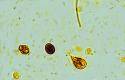
The Giardia trophozoite - which is the active stage of the organism - inhabits the small intestine
of the dog. It attaches to the cells of the intestine with its adhesive disc and rapidly divides to produce a whole population
of trophozoites. As they detach they may be swept down the intestine. If intestinal flow is fast then they may appear in the
faeces. However, if they have time, they will develop into the inactive, more durable, cyst form of the organism and these
will be passed in the faeces. The cyst is more able to survive in the environment than the trophozoite, which is very fragile.
How do Giardia cause disease in dogs?
Like all infectious agents, in order to cause disease Giardia depaends on being able to overcome
the dog's defence against infection, either by its virulence or by the number of the organisms becoming established. It has
been observed that as few as 10 cycsts can cause disease in humans. Different anaimals may respond to infection in different
ways, which may be due to different strains of the sam Giardia population, with varying levels of pathogenicity.
Another explanation for observed differences in the host response to infection is that protective immunity with age and/or
exposure. This may be temporarily lost if the animal is stressed or immunosuppressed, for example with corticosteroid treatment.
What is the source of infection for dogs?
The original source of an outbreak may be cysts in contaminated water or the environment. In addition,
infected dogs which may be either carriers (ie: show no clinical signs but continue to harbour infection and pass cysts into
the environment) or dogs that have diarrhoea associated with infection may act as the source. Surveys have shown that about
14% of the adult dog population and over 30% of dogs under one year of age were infected. Once passed, the cysts can survive
in cold water for several months.
The cysts are infective as soon as they are passed, unlike other parasites where a lag period is necessary
before the organism is infective. The most common route of infection is faeco-oral. For example, dogs may accidentaly eat
cysts as they lick around theenvironment or lick other dogs' coats (particularly if the other dog has diarrhoea). Another
major source of infection in human cases is drinking contaminated water. Once eaten, the cyst breaks open in the animals'
intestine and releases two new trophozoites to initiate infection. If a dog is left in a dirty environment it may act as its
own source of further infectionas it eats cysts passed in its own faeces.
What are the clinical signs associated with infection?
The trophozoites divide to produce a large population, then they begin to interfere with the absorption
of food, so faeces from affected animals are typically light coloured, greasy and soft. These signs, together with the beginning
of cyst shedding, begin abou tone week post-infection. There may be additional signs of large intestinal irritation, such
as straining and mucus in the faeces, even though the Giardia do not colonise the large intestine. Usually the blood
picture of affected animals is normal, though occasionally there is a slight increase in the number of eosinophils (one of
several types of white blood cells) and mild anaemia. Without treatment, the condition may continue, either chronically or
intermittently, for weeks or months.
How can infection be diagnosed?
Diagnosis is based on demonstration of the infection and the elimination of other possible causes of
diarrhoea (eg: Salmonella or Campylobacter), Giardia cysts may be observed directly in faecal samples or
indirectly using an elisa technique. Direct examination of faeces, using zinc sulphate centrifugal flotation. followed by
staining the supernatant with Lugol's iodine, has been found to be upto 70% effective at detecting infection from a single
faecal sample. The cyst output is very variable from day to day so the detection rate may be improved by pooling faecal samples
collected over three days. Faecal examination is the cheapest method but is time consuming and requires an experienced technician
for reliable results.
The elisa technique requires a kit and some method of reading a colour change or production of flourescence.
Studies examining the reliability of some immunoflourescent kits have found them to be over 90% accurate, with relatively
few false negatives or false postives. However, the tests are costly and probably only wothwhile where there are alarge number
of samples to be processed and a technician who is familiar with carrying out elisas.
How can infection be treated?
Infection may be treated using one of a number of drugs. Unfortunately there is no treatment licenced
for the control of giardias in dogs, though fenbendazole (Panacur, Hoechst Animal Health) is licenced for treatment of worms
in dogs. Whatever treatment is chosen, it is very unlikely to eliminate 100% of the infection in all dogs.
Adaptations that may be made to try to improve the success rate of a treatment regime include extending
the duration and dose of the treatment. Care must obviously be taken with this approach to make sure that an adequate safety
margin is always maintained. Another approach is to retreat after an interval of one week. Alternatively, repeat faecal samples
may be collected one week after the treatment and dogs which are still passing cysts can be identified and treated. It should
be recognised that, when treating a large number of dogs, whichever of these treatment strategies is adopted, there may be
one or two dogs that remain as carriers of infection that will act as a potential sources of infection in future.
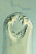
Cherry Eye
When the tear gland of the third eyelid pops out of position, it protrudes from behind the eyelid
as a reddish mass. This prolapsed tear gland condition is commonly referred to as "cherry eye". The problem is seen primarily
in young dogs, including the Cocker Spaniel, Lhasa Apso, Shih-Tzu, Poodle, Beagle, and Bulldog. It's also seen sometimes in
certain cat breeds including the Burmese.
Despite its appearance, cherry eye itself is not a painful condition. However,
the longer the tear gland is exposed, the more likely it will come irritated and inflamed. If the patient rubs at the eye,
it could cause the gland to bleed or become infected. Furthermore, the function of the tear gland could become compromised
if the gland is exposed for long periods of time.
To correct cherry eye, surgical REPLACEMENT of the gland is necessary.
This treatment is superior to a somewhat older technique of surgically REMOVING the gland. The gland of the third eyelid plays
an important role in maintaining normal tear production. We now know that dogs who have had the tear gland removed are predisposed
to developing Dry Eye Syndrome later in life. Dry Eye Syndrome is uncomfortable for the patient, and requires the owner to
administer topical medications several times a day for the remainder of the patient's life. To avoid this condition, it is
preferable to tuck the tear gland back inside the third eyelid, where it can continue to function normally.
Under Construction...
|
|
|
|
|
|